. 2020 Oct 22;9:e58593. doi: 10.7554/eLife.58593

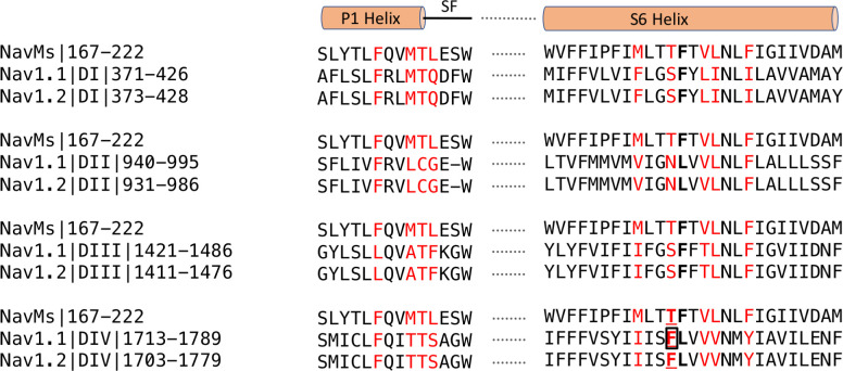
Figure 6. Sequence alignments of CBD-binding site regions of NavMs With corresponding regions in hNav1.1 and hNav1.2.

In red are the CBD-binding residues (within 3.9 Å of the compound) in NavMs and the equivalent residues in hNav1.1 and hNav1.2. The bold black F indicates the site of the NavMs F208L mutant used in these studies. It was changed from F to L in NavMsL because in half of the human Nav domains it is an F and in the other half it is an L (both indicated in bold black in the other sequences). However, as shown in Figure 6—figure supplement 2, the residue type present at this site makes essentially no difference in the structure. Residues located in the binding site are found within the P1 pore helix, the selectivity filter loop, and the S6 helix. The residue, which when mutated to alanine in hNav1.1 reduces the binding affinity of CBD (Ghovanloo et al., 2018), is boxed. This corresponds to T207 in NavMs, which is the residue that was mutated to alanine in the electrophysiology characterisations in the present study. The sequence alignment was carried out using Clustal Omega (Sievers et al., 2011) and annotated manually. 

Figure 6.

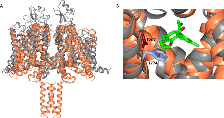
Figure 6—figure supplement 1. Alignment of the NavMs-CBD structure (coral) with a homology model of hNav1.1 (grey).

Figure 6—figure supplement 1.

The homology model was created in SWISS-MODEL (Waterhouse et al., 2009) using hNav1.2 (PDB code 6J8E) as a template. (A) Overall structural alignment. (RMSD = 3.34 Å for the transmembrane regions). (B) Detail of this overlay showing the location of CBD (green and red stick depiction) in the region of NavMs residue T207 (light blue stick representation); this residue corresponds with residue F1774 in hNav1.1, which is a residue that has been identified as being in the local anaesthetic binding site, and was the residue that was mutated in electrophysiology experiments (Ghovanloo et al., 2018), which showed the effects of CBD inhibition on hNav1.2.
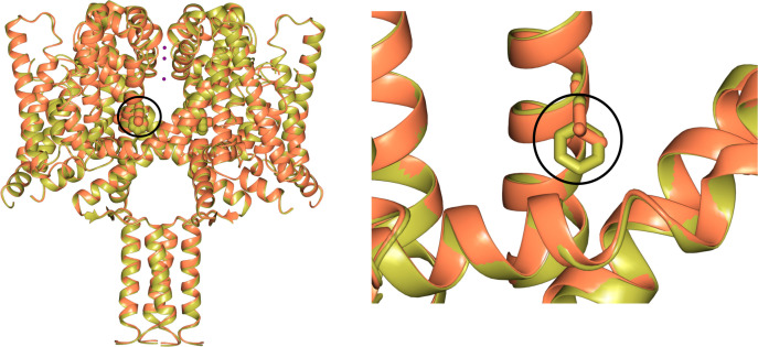
Figure 6—figure supplement 2. Comparisons of wild type NavMs (gold) [PDB ID 5HVX] and the NavMsL mutant (coral) [PDB ID 6YZ2] structures.

Figure 6—figure supplement 2.

These figures show (left) the close overall similarities of the structures, and (right) the detailed region surrounding mutated residue F208L (which is circled in both panels, and is indicated in bold in the sequence alignment shown in Figure 6).