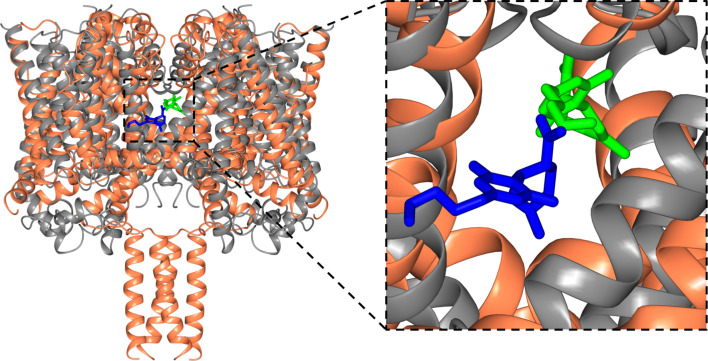
Figure 8. Structural alignments of the NavMs-CBD (coral ribbons) crystal structure and the TRPV2-CBD cryo-EM structure [PDB ID 6U88] (grey ribbons).
(Left) Overall alignment of the structures. The TRPV2 structure was trimmed to remove the extramembranous regions for clarity. The RMSD of the alignment is 4.2 Å. The CBD in NavMs is shown in green and that in TRPV2 is in blue. The CBD site in NavMs appears to be located further into the fenestration than it is in TRPV2. (Right) Detailed view of the CBD sites, highlighting the similarity and differences in orientation and location in the two channel types.

