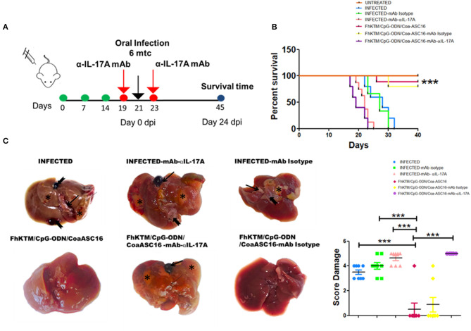
Figure 7.
IL-17A neutralization decreased the efficacy of FhKTM/CpG-ODN/Coa-ASC16 vaccination. (A) Mice were administered with anti-IL-17A antibody and the corresponding isotype control (250 μg i.p) 2 days before and after infections. (B) Kaplan–Meier graphs show survival curves for those mice immunized three times with FhKTM/CpG-ODN/Coa-ASC16 or PBS. IL-17A-mAb and corresponding isotype control (mAb Isotype) were administered 2 days both before and after infection. dpi: days post infection. (C) Left: infected liver showing marked enlargement with distended gallbladder (thick arrow), areas of fibrosis extending (asterix), and larvae on the parenchyma surface (notched arrow). FhKTM/CpG-ODN/Coa-ASC16: liver showing normal size with no observed lesion except in 1 out of 8 mice. Infected-mAb-IL-17A: the liver showing marked severe macroscopic damage (thin arrow) and presented irregular surface denoting hepatic fibrosis (asterix). The presence of larvae on the surface can be seen (notched arrow). FhKTM/CpG-ODN/Coa-ASC16 -mAb-IL-17A: The hepatic parenchyma is severely reduced, firm, and presents an irregular surface (thin arrow) denoting hepatic fibrosis (asterix). Abnormal gross appearance of the distended gallbladder with presence of blood inside (thick arrow). infected-mAb Isotype: the liver is smaller than normal and presents irregular surface tissue denoting hypertrophied (thin arrow) and replaced with fibrosis (asterix) and a fluke released after the rupture of the gallbladder (notched arrow). FhKTM/CpG-ODN/Coa-ASC16-mAb Isotype: liver showing no apparent gross pathology except for in 2 out of 8 mice. Right, the score damage was assessed by the extensión of liver damaged (score range 0 to 5). The data shown are pooled from two independent experiments with total (n = 5–8 per group). The statistical differences among survival curves were calculated by using Mantel–Cox test. ***p < 0.001.

