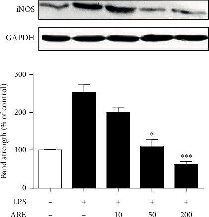
Figure 6.

Suppressive effect of ARE on the LPS-stimulated-inducible iNOS production in HaCaT keratinocytes. After the 5.0 × 10⁵ HaCaT cells were preincubated with the varying concentrations (0, 10, 50, and 200 μg/mL) of ARE, the cells were treated with 1 μg/mL LPS for 24 h. iNOS in cellular lysates was detected using western blotting analysis. GAPDH was used as a protein loading control. In the lower panel, the relative band strength, expressed as % of control, was determined with densitometry using the ImageJ software which can be downloaded from the NIH website. ∗P < 0.05; ∗∗∗P < 0.001 versus the LPS only.
