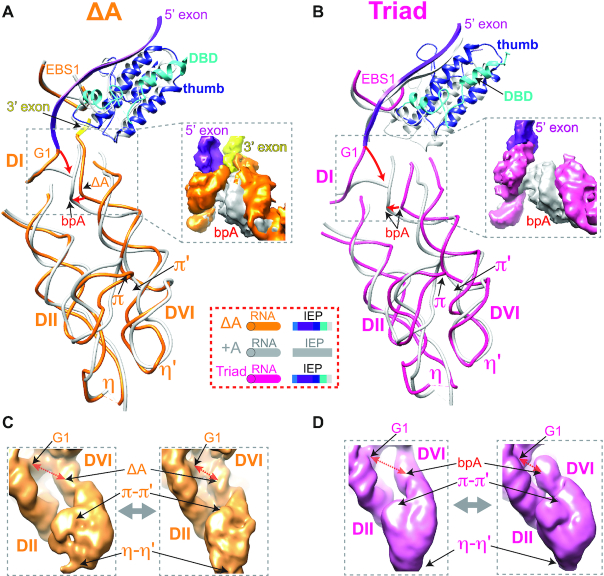
Figure 6.
Pre-catalytic configurations of the DII-DVI complex. (A, B) Structures of DII–DVI region in the ΔA (A) and the Triad (B) RNPs are compared to the +A RNP (PDB: 5G2X). RNAs of ΔA, +A and Triad are in orange, grey and dark pink, respectively. +A IEP is in grey and the precursors’ IEPs are shown in their corresponding colors. ‘bpA’ denotes the branch point adenosine and the orange ‘ΔA’ with arrow pointing to the branch point denotes adenosine deletion in the ΔA RNP. π-π′ and η-η′ motifs are indicated. The 5′ exons of the ΔA and Triad RNPs are in magenta. The 3′ exon, which is not visible in the Triad RNP, is in yellow of the ΔA RNP. (C, D) Intrinsic RNA flexibility of the DVI-DII pair as shown in Supplementary Movie 4. 3D variability analysis of ΔA (C) and Triad (D) RNPs identified conformers of DII-DVI region for each of the precursor RNPs, with representative conformations having the branch site (ΔA or bpA) away from or closer to the 5′ end of the intron (G1).

