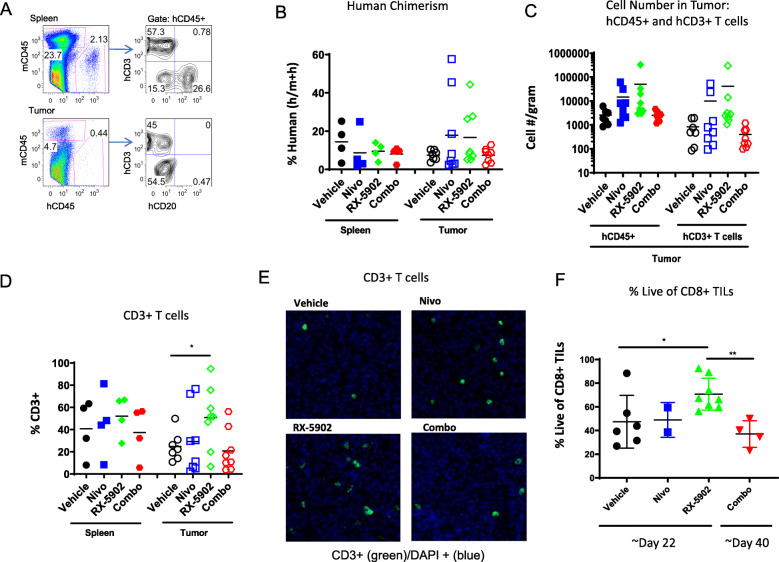
Fig. 3.
Human immune subsets in spleens and tumors of hu-CB-BRGS MDA-MB-231 bearing mice at the end of study. a Flow cytometry panel for identification of mouse (mCD45) and human (mCD45) hematopoietic cells, and T (CD3) and B (CD20) cells among the hCD45+ population (right), in the spleen (top) and tumor (bottom). b Percentage of human (hCD45+ of (mCD45+ + hCD45+)) cells in the spleens and tumors. c T cell (CD3+) numbers and frequencies and d among the hCD45+ cells. e Representative fields from tumors collected at end of study from the indicated treatment arms stained with anti-CD3 antibodies (green) and DAPI and imaged using the Vectra 3.0 system. f Frequency of live cells among CD8+ human (hCD45+) cells in tumors. Each dot represents data from an individual spleen, bone marrow or tumor from mice receiving either no drug (Vehicle), nivolumab alone (Nivo), RX-5902 alone, or combination of nivolumab and RX-5902 (Combo) treatment. Lines represent arithmetic means. p-value: * < 0.05

