
Figure 1.
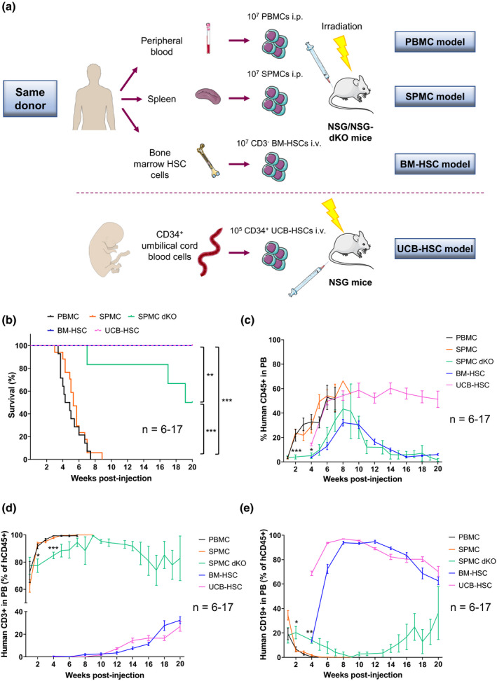
Distinct survival and human reconstitution in the different humanised mouse models. (a) Schematic representation of the generation of different humanised mouse models. To prevent donor‐related variations, PBMCs, SPMCs and BM‐HSCs used for reconstitution of NSG mice, were isolated from the same three adult donors (Donors 1–3). For the SPMC dKO model, NSG‐dKO mice were humanised with SPMCs from Donor 3. For the UCB‐HSC model, NSG mice were reconstituted with UCB‐HSCs cells from Donor 4. (b) Kaplan–Meier plot showing overall survival of the different humanised mouse models. Mice were sacrificed at 20 weeks or earlier if they showed clinical signs of GvHD. (c) Levels of hCD45+ cells circulating in PB of mice at the indicated time points. (d) Levels of hCD3+ T cells and (e) hCD19+ B cells circulating in PB of mice. Data in the PBMC (n = 14), SPMC (n = 17) and BM‐HSC (n = 13) models are representative of three independent experiments with Donors 1–3 in each model. Data in the SPMC dKO model (n = 6) are representative of one experiment with Donor 3. Data in the UCB‐HSC (n = 7) are representative of one experiment with Donor 4. Data are mean ± SEM. The Mantel–Cox test was used to assess statistical differences between models in b, and a two‐way ANOVA to assess differences at each time point between models in c–e; *P < 0.05, **P < 0.01, ***P < 0.001. BM‐HSC, bone marrow haematopoietic stem cells; i.p., intraperitoneally; i.v., intravenously; NSG, NOD scid gamma; NSG‐dKO, NOD scid gamma double knockout; PB, peripheral blood; PBMC, peripheral blood mononuclear cells; SPMC dKO, SPMC model in NSG‐dKO mice; SPMC, spleen mononuclear cells; UCB‐HSC, umbilical cord blood haematopoietic stem cells.