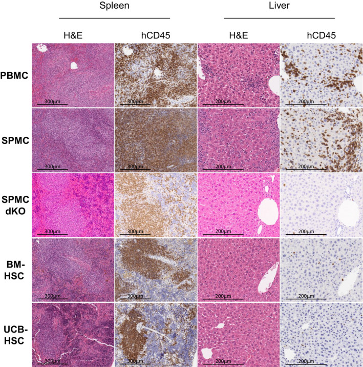
Figure 2.

Histological comparison of human immune cell infiltration and inflammation levels in mouse tissue. Representative images of the H&E and hCD45 staining on the spleen and liver of one mouse in each model, reconstituted with Donor 3 (PBMC, SPMC, SPMC dKO and BM‐HSC models) and Donor 4 (UCB‐HSC model). PBMC and SPMC mice shown were sacrificed at week 5 and 6, after GvHD development. SPMC dKO, BM‐HSC and UCB‐HSC mice shown were sacrificed at study end at week 20. Magnification: ×400 for the liver and ×200 for the spleen. BM‐HSC, bone marrow haematopoietic stem cells; H&E, haematoxylin and eosin; PBMC, peripheral blood mononuclear cells; SPMC dKO, SPMC model in NSG double knockout mice; SPMC, spleen mononuclear cells; UCB‐HSC, umbilical cord blood haematopoietic stem cells.
