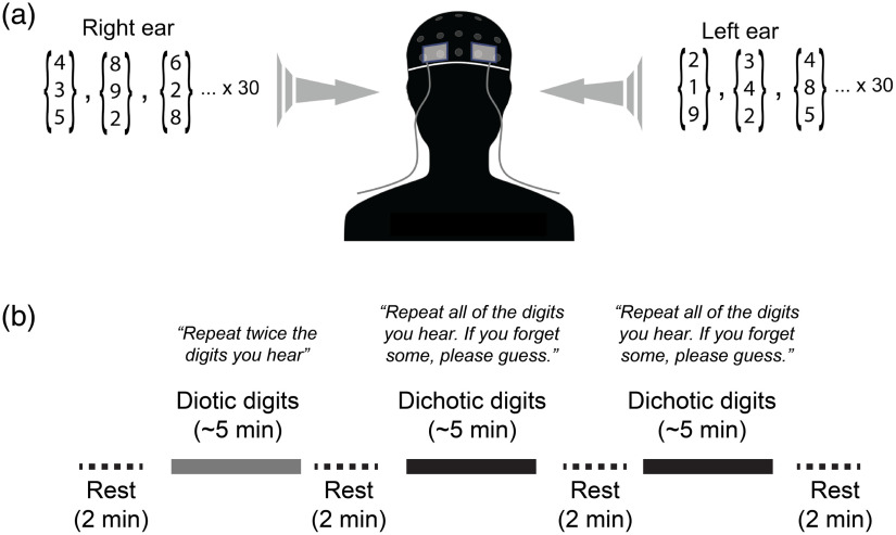
Fig. 1.
Experimental apparatus and protocol. (a) Optical probes embedded in flexible elastomer were placed at locations AF7 and AF8 (10-10 international electrode placement system) and secured under a neoprene EEG cap. The three-digit arrays depict the groups of three spoken digits that were selected randomly and delivered to each ear through calibrated insert earphones during dichotic listening tasks. (b) A block paradigm timeline indicating the timing of test blocks and rest periods. Specific paraphrased instructions are shown above each task block.

