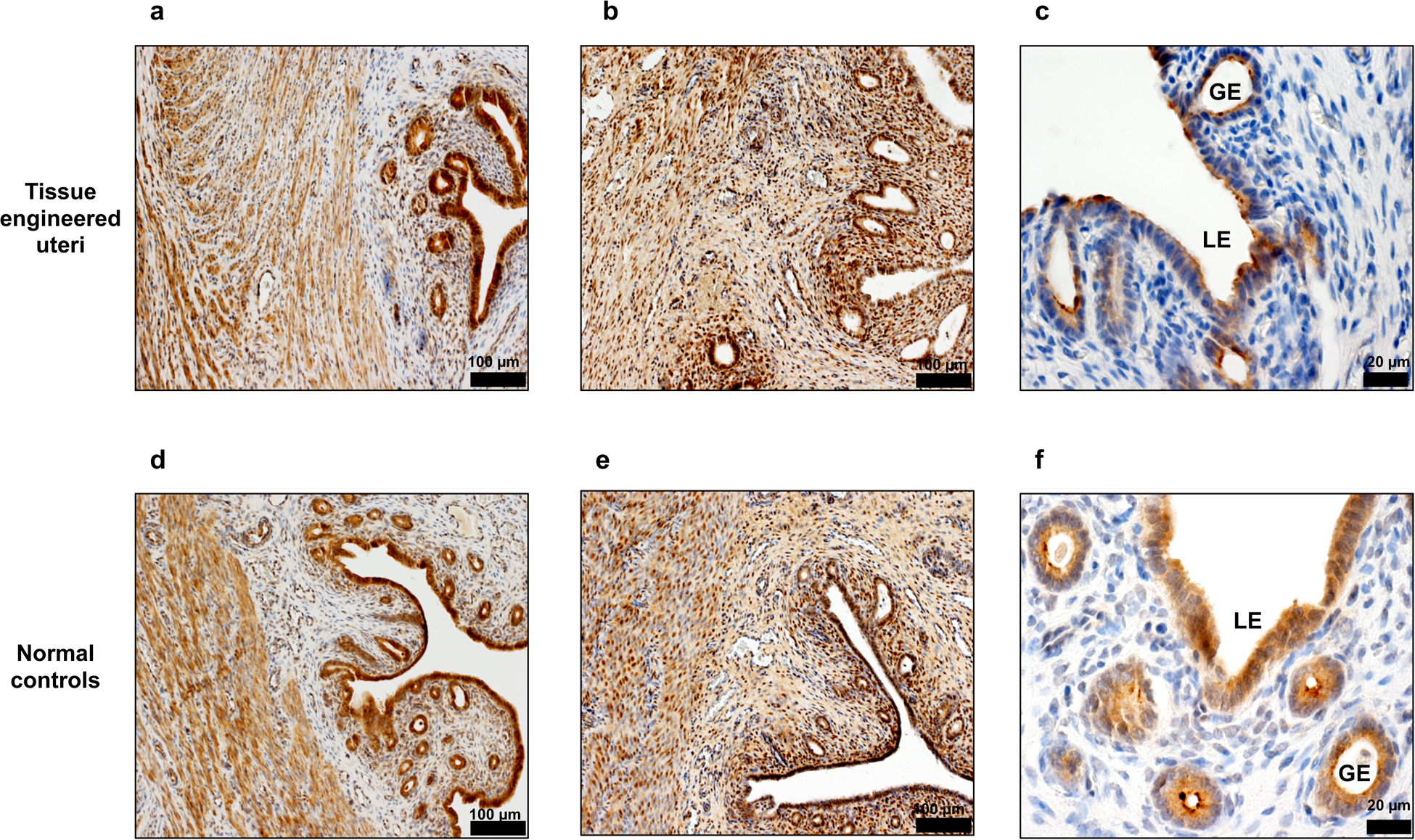
Figure 4.

Immunohistochemical staining of functional markers in the tissue engineered uteri at 6 months post implantation. (a,d) Estrogen receptor alpha positive cells in the endometrium and myometrium layers. Scale bar: 100 μm (b,e) Progesterone receptor expression in epithelial and stromal cells. Scale bar: 100 μm (c,f) Detection of uteroglobin expression showed the presence of secretory glands structure in the engineered uteri. Data shown are representative images from n = 3 animals; experiments were repeated independently three times with similar results. Scale bar: 20 μm. LE: luminal epithelium. GE: glandular epithelium.
