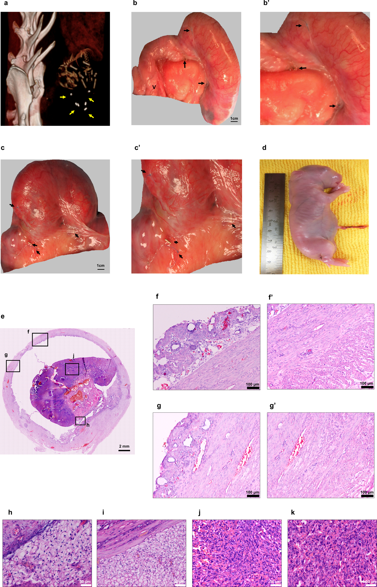
Figure 5.

Fetal development site identification at day 29–30 post mating in a single-horn pregnant uterus. (a) Computed tomography image of a fetus within the tissue engineered uterus, demarcated by the titanium clips. (b-c) Pregnancies in the tissue engineered uteri group at birth. Scale bar: 1 cm. (b’-c’) Titanium clips and marking sutures indicated that fetal development occurred within the bioengineered segment. (d) Gross appearance of a newborn from a tissue engineered uterus. (e) H&E stained cross-section of the pregnant bioengineered uterus. Scale bar: 2 mm (f, g) Micrographs of the pregnant engineered uterine wall revealed structural integrity of the endometrial and myometrial layers (f’, g’). Scale bar: 100 μm (h, i) Decidua. (h) Decidua zone in the tissue engineered uteri group; (i) decidua zone in normal controls. (j, k) Rabbit placenta. (j) Labyrinth zone in the tissue engineered uteri group; (k) labyrinth zone in normal controls. Scale bar: 50 μm. Yellow arrows indicate titanium clips; black arrows indicate the titanium clips and non-absorbable sutures at the margins of the engrafted tissue. Data shown are representative images from n = 4 animals; experiments were repeated independently three times with similar results. V: vagina.
