
Fig. 1.
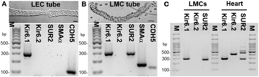
A) An image of an isolated LEC tube dissociated from a mouse popliteal lymphatic vessel (top), and PCR results (bottom) showing message for Kir6.1 and CDH5 (VE-cadherin) but not Kir6.2, SUR2 or SMA α-actin; B) An image of LMCs remaining after expulsion of an LEC tube (top), and PCR results (bottom) showing message for Kir6.1, SUR2 and SMA α-actin, with some contamination from CDH5. C) PCR results from LMCs purified by FACS using 4 pooled, inguinal-axillary lymphatic vessels from a tamoxifen-treated SMMHC-CreERT2;Rosa26mTmG mouse, showing message for Kir6.1 and SUR2, but not Kir6.2; heart was used as a positive control. Kir6.1 (Kcnj8) = 301 bp; Kir6.2 (Kcnj11) = 430 bp; SUR2A (Abcc9) = 465 bp; SUR2B (Abcc9) = 289 bp; SMA = 146 bp; CDH5 = 116 bp. PCR results are representative of 3 experiments. Calibration bars = 50 μm.