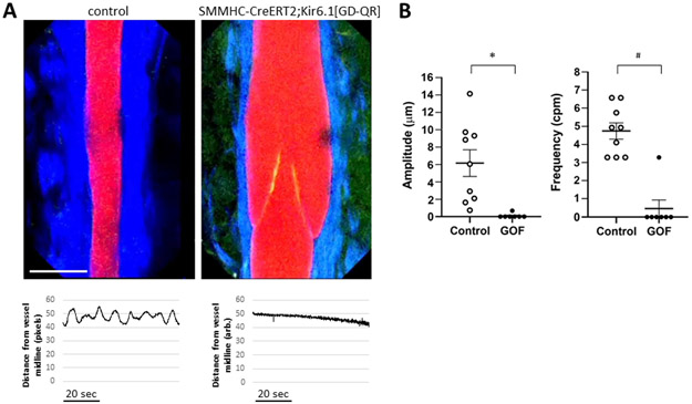
Fig. 9.
A) In vivo images of popliteal lymphatic collectors from control mice (left) and TMX-treated SMMHC-CreERT2;Kir6.1[GD-QR] mice taken at the minimum diameter of the lymphatic contraction cycle. Red= lectin; Green = GFP, Blue = 2nd harmonic (collagen); calibration bar = 80 μm. An intraluminal valve (with GFP+ leaflets) is visible in the latter. Below each image is an example of the diameter change from the vessel midpoint over a 1.2 min period, measured from the lectin channel; it shows that cyclical diameter changes in the control vessel and absence of diameter change in the TMX-treated SMMHC-CreERT2;Kir6.1[GD-QR] vessel. B) Summary of amplitude and frequency of diameter fluctuations in control (n=9) and GOF (SMMHC-CreERT2;Kir6.1[GD-QR]) (n=7) vessels. * = p< 0.0002; # = p<0.0007.

