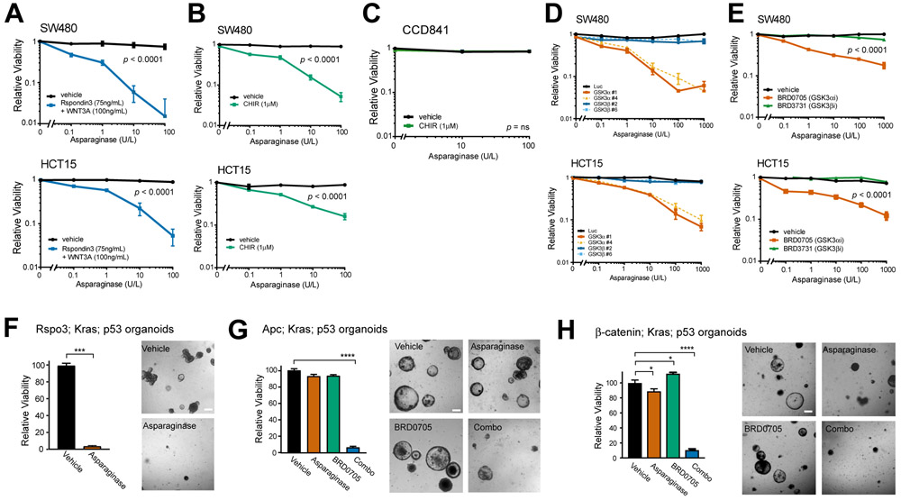
Figure 1. Activation of WNT signaling upstream of GSK3 induces asparaginase hypersensitivity.
A, SW480 and HCT15 cells were treated with the indicated doses of asparaginase for 10 days, in the presence of recombinant Rspondin3 (75 ng/ml) and WNT3a (100 ng/ml) or vehicle. The number of viable cells was counted by trypan blue vital dye staining, and all cell counts were normalized to those in vehicle-treated cells (no asparaginase, Rspo3 or WNT3a). Two-way ANOVA was performed for each cell line and included interaction terms between asparaginase doses and WNT ligands. The p value for the main effect of WNT ligands vs. vehicle is presented in each plot and the interaction terms were overall significant (p < 0.0001).
B, SW480 and HCT15 cells were treated with the GSK3 inhibitor CHIR99021 (CHIR, 1μM) or vehicle, together with the indicated doses of asparaginase for 10 days. Viability was assessed as in (A). Statistical significance was calculated using a two-way ANOVA and included interaction terms between asparaginase doses and GSK3 inhibitor. The p value for the main effect of WNT ligands vs. vehicle is presented in each plot and the interaction terms were significant (p < 0.0001).
C, CCD841 cells derived from normal human colonic epithelium were treated with CHIR99021 (1 μM) or vehicle, together with the indicated doses of asparaginase for 10 days, and viable cell counts were assessed as described in (A). Two-way ANOVA was performed for each cell line and included interaction terms between asparaginase dose and GSK3 inhibitor. The interaction terms were not significant (p = ns).
D, SW480 and HCT15 cells were transduced with the indicated shRNAs and then treated with the indicated doses of asparaginase. Viability was assessed after 10 days of treatment by counting viable cells. All cell counts were normalized to those in shLuc-transduced, vehicle-treated controls.
E, The indicated cell lines were treated with vehicle, the GSK3α-selective inhibitor BRD0705 (1 μM), or the GSK3β-selective inhibitor BRD3731 (1 μM), in the presence of the indicated doses of asparaginase for 10 days. Viability was assessed as in (A). Two-way ANOVA was performed and included interaction terms between asparaginase dose and type of GSK3 inhibitor type. The p value for the main effect of GSK3 inhibitor type is presented.
F, Mouse intestinal organoids expressing an endogenous Rspo3 fusion in addition to p53 loss-of-function and an activating KrasG12D mutations were cultured in basal medium (which lacks WNT/R-spondin supplementation) and treated with vehicle or asparaginase (100 U/L) for 10 days. Viability was assessed by counting viable organoids using an Axio Imager A1 microscope. Images were taken from a representative of three experiments, and statistical significance was calculated using a two-sided Welch t-test. Scale bar, 100 μm.
G, Apc-deficient organoids with mutations of p53 and Kras were cultured in basal medium and treated with vehicle, asparaginase (100 U/L), BRD0705 (1 μM) or combo (100 U/L asparaginase + 1 μM BRD0705) for 10 days. Viability was assessed as in (F). Images were taken from a representative of three experiment. Scale bar, 100 μm. Differences between groups were analyzed using a one-way ANOVA with Dunnett’s adjustment for multiple comparisons, using the vehicle group as the reference group.
H, Organoids with a β-catenin activating mutation, as well as Kras and p53 mutations, were cultured in basal medium, treated and analyzed as in (G).

