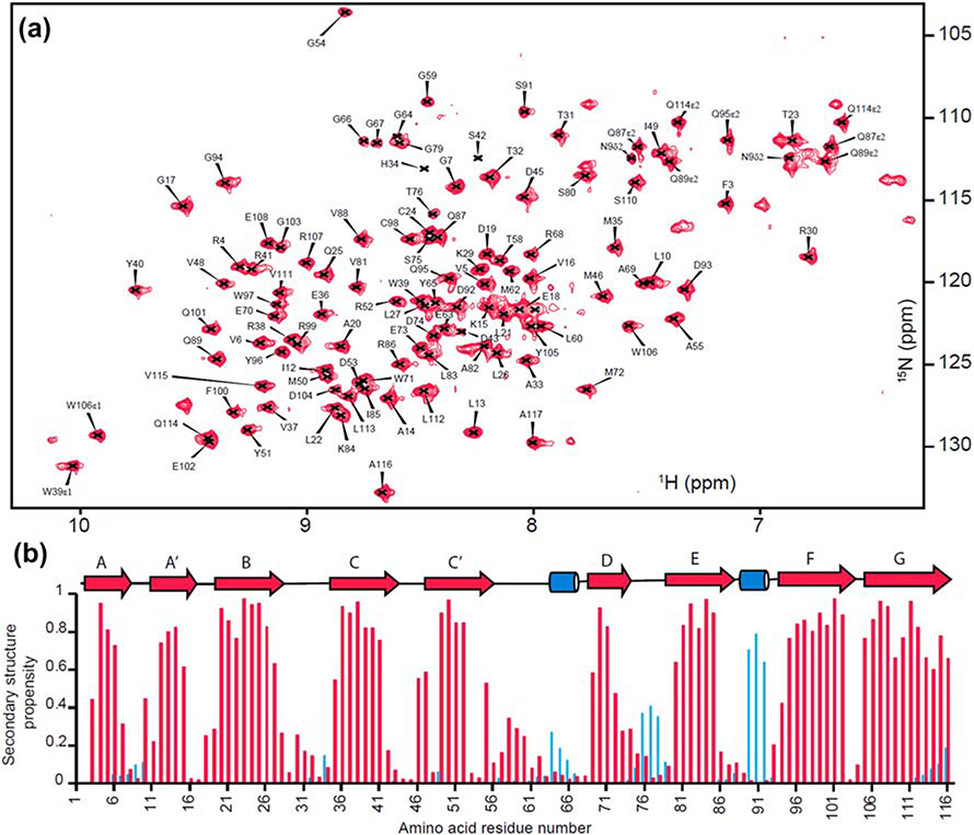
Figure 3: Backbone assignment and secondary structure propensity of BTNL2 IgV domain.
A. Assigned 15N-1H HSQC spectra of mouse BTNL2 IgV domain. B. Secondary structure propensity calculated from backbone chemical shifts using MICS. β-strands and α-helices are colored in red and blue, respectively, and also shown as arrows and cylinders, respectively.

