FIG 1.

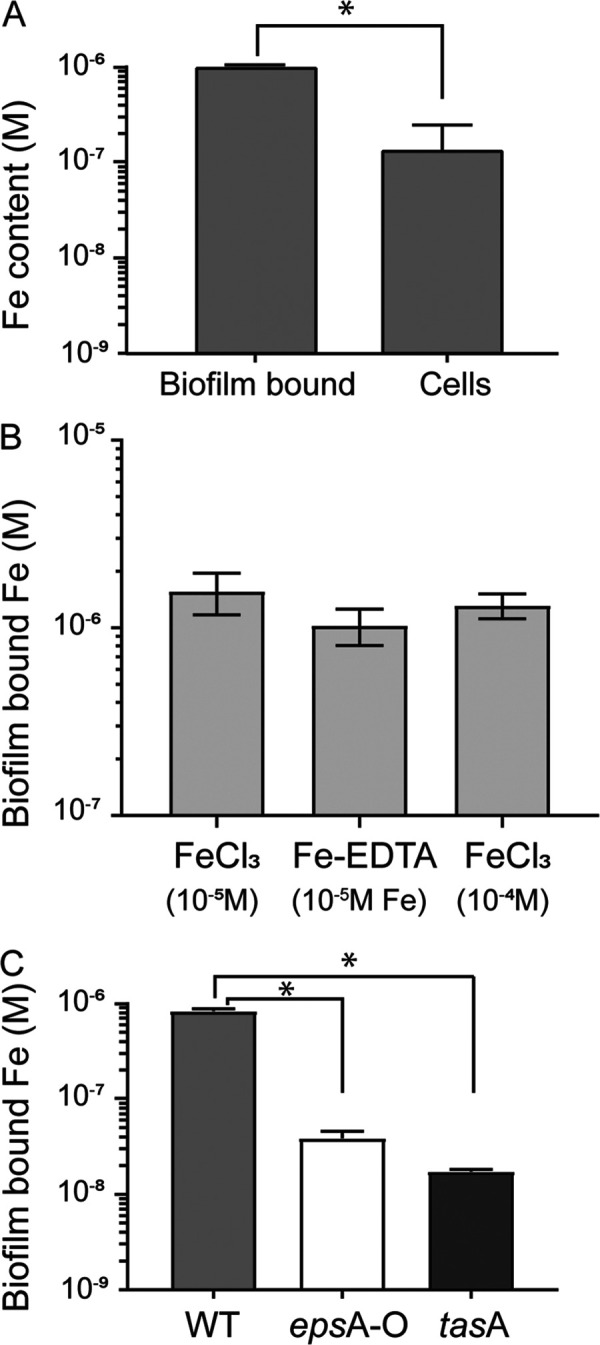
B. subtilis biofilm accumulates Fe. (A) Fe content (in molar) measured in B. subtilis cells and biofilm matrix after 22 h of growth at 30°C in MSgg supplemented with 10−4 M FeCl3 (an asterisk indicates significant difference by t test, P < 0.001). (B) Fe content (in molar) measured in B. subtilis cell biofilm matrix following 28 h of growth at 30°C in MSgg supplemented with Fe provided as FeCl3 (10−5 and 10−4 M) and Fe-EDTA (10−5 M FeCl3 + 10−4 M EDTA). Error bars are standard deviations. (C) Fe content of biofilms formed by the wild type (light gray bars), epsA-O (no exopolysaccharides, white bars), and tasA (no TasA fibers, dark gray bars) cells measured after 22 h of growth at 30°C in MSgg supplemented with 10−4 M FeCl3 (an asterisk indicates significant differences by ANOVA with post hoc Tukey’s test, P < 0.001).
