
Figure 4: sFRP2 treatment overcomes response to anti-VEGF.
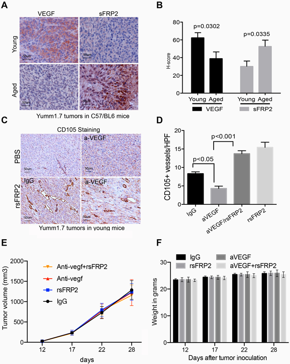
A. Representative immunohistochemistry of VEGF and sFRP2 staining in tumors in young and aged mice; B. Quantification of staining in A, based on the H-score calculated by percent of tumor positive, and intensity of stain. C. Representative CD105 immunohistochemistry of primary Yumm 1.7 mcherry murine tumors in young C57BL/6 mice treated with PBS or rsFRP2, in the presence of the anti-VEGFA antibody, or an IgG control. D. Quantification of staining in C. E. Yumm 1.7 cells were implanted in mice and then mice were treated with antibody treatments as indicated, and tumor growth was measured. F. Mouse weight in grams after indicated treatments, beginning 12 days after tumor inoculation.