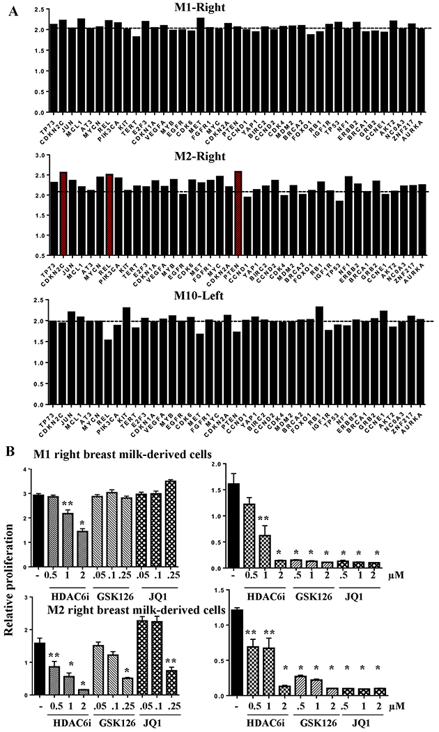
Fig. 7: Sensitivity of right breast milk-derived cells of donors M1 and M2 to targeted therapies.

A) CNV analysis of DNA from breast milk-derived cells of M1, M2 and M10. CNVs in DNA from breast milk-derived cells of right of M1, right of M2 and left of M10 were determined by comparing to DNAs from breast milk-derived cells of left of M1, left of M2 and right of M10. Horizontal line indicates normal copy number value of 2. Genes with numbers close to 2.6 or above are considered amplified (indicated in red in case of M2-Right). Raw values for all 87 genes are provided in Table S2. B) Cells were treated with indicated drugs for five days and cell proliferation was measured using bromodeoxyuridine incorporation-ELISA. Results of two independent experiments at different concentrations of GSK126 and JQ1 are shown. Data shown are average and standard errors of six technical replicates. *p<0.001. **<0.02, untreated versus drug treated cells.
