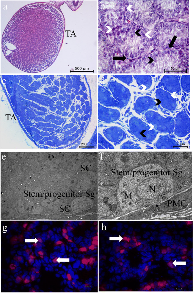
Fig. 1.
Light (a–d) and electron (e, f) micrographs of a 6-day-old C57/BL-6 mouse testis The low a and b high power micrographs of paraffin sections exhibit testicular lobules consisting of seminiferous cords and the tunica albuginea (TA) that surrounds the testis. Seminiferous cords are surrounded by monolayer of peritubular myoid cells (black arrows) and the spermatogonia (white arrowheads) are located in the cords. c and d are the semi-thin plastic section micrographs clearly showing the presence of the basally located large, bright spermatogonia (white arrowheads) and Sertoli cells (black arrowheads) arranged perpendicularly to the basement membrane inside the seminiferous cords. e and f present the Sertoli cells (SC) and the stem/progenitor spermatogonia at the ultrastructural level within the cords. Note the high amount of mitochondria (M) in the cytoplasm and small heterochromatin clumps inside the nuclei (N) of the stem/progenitor spermatogonia and peritubular myoid cell (PMC) at high magnification. g and h are the frozen sections showing the PLZF (+) stem/progenitor spermatogonia (white arrowheads) in the seminiferous cords by IF. a, b Hematoxylin eosin × 100 and × 600. c, d Methylene blue azure II × 100 and × 400. e, f Uranyl acetate–lead citrate × 4000 and × 20000. g, h IF × 1000

