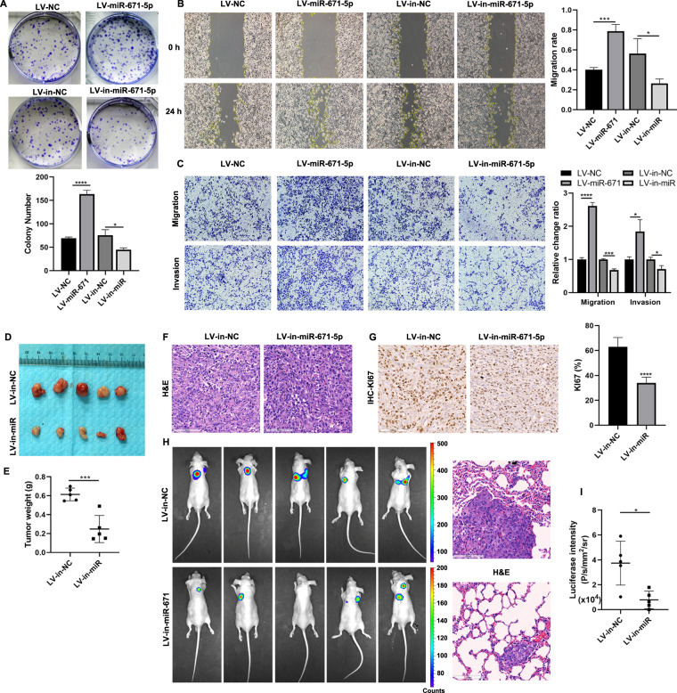
Fig. 3. MiR-671 facilitated PCa cells proliferation, migration, and invasion in vitro and in vivo.
Colony formation assays (A), wound healing assays (B), and Transwell assays (C) were performed to assess the proliferation, migration, and invasion ability in miR-671 knockdown or overexpressing PC-3 cells. D In vivo PCa tumor formation in the xenograft male nude mouse models. E Final tumor weights were measured. F The xenograft tumor tissues were stained with H&E. G KI67 IHC staining in xenografts with LV-in-miR-671 cells or LV-in-NC cells. (H-left and I) The tail vein xenograft model was used to investigate the effects of miR-671 on PCa metastasis in vivo, and lung colonization ability was measured by bioluminescence imaging. (H, right) The lung sections were stained with H&E. Magnification, ×200. Scale bars, 100 μm. The data were presented as means ± SD from three biological replicates. *P < 0.05; **P < 0.01; ***P < 0.001; ****P < 0.0001; Student’s t-test.

