Figure 1.
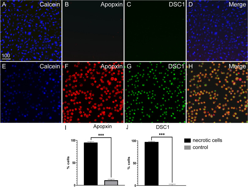
Heat exposure induces necrosis of McCoyB cells. The cells were either left untreated or induced to undergo necrosis by heat exposure at 55 °C for 30 min, after which cells were stained and transferred to 96-well plates for imaging. All cells were labelled with cytocalcein (live cell stain, blue), PS sensory dye (Apopxin, red) and membrane-impermeable DNA nuclear dye (DCS1, green). Example images of (A–D) untreated control cells and (E–H) heat-treated cells. (I) Percentages of cells displaying PS. (J) Percentages of dead cells (cells labelled with DSC1). PS- and DCS1-positive cells were automatically counted using Nikon Elements software. ***P ≤ 0.0001 (unpaired t-test with Welch’s correction). Data represents mean ± SEM (3 biological × 3 technical replicates of ~ 400 cells × 4 FOV). Scale bar: 100 µm.

