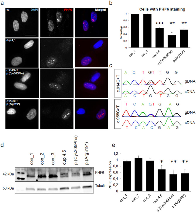
Figure 2.
Variants in PHF6 in female individuals lead to a loss of protein. In fibroblasts of female individuals with variants in PHF6, only the wildtype PHF6 allele was expressed. (a) Immunofluorescence of fibroblasts of three individuals with either a duplication of exons 4 and 5, a missense variant c.914G>T (p.(Cys305Phe), or a truncating variant c.955C>T (p.(Arg319*) and one control individual. In the control, PHF6 expression could be observed in all fibroblast cells. In affected individuals, expression of PHF6 was present in only 50% of cells. Scale bar represents 20 µm. Cells were stained with rabbit polyclonal anti-PHF6 antibody (HPA001023, Sigma-Aldrich, 1:50). (b) Numerical analysis of fibroblasts with variants in PHF6 and controls. Affected individuals showed expression of PHF6 in 30–50% of cells, respectively. For each individual, at least 63 cells were evaluated. (c) Sanger sequencing of genomic DNA (gDNA) and of cDNA from RNA of affected individuals. In the individuals with a missense or a truncating variant respectively, both wildtype and mutant allele could be observed on gDNA level. On cDNA level, only the wildtype allele was present, thus indicating nonsense mediated mRNA decay. (d) Exemplary western blot from fibroblasts of control and affected individuals stained for PHF6 (Santa Cruz Biotechnology, sc-365237, 1:500) and Tubulin (ab7291, abcam, 1:10,000) as a housekeeping gene. PHF6 levels in patient fibroblasts were decreased. Grouped blots were cropped from different blots. Uncropped blots can be found in Supplementary Fig. S2. (e) Graphical analysis of three western blot replicates indicating 50%-70% of remaining PHF6 protein in patient fibroblasts. Error bars depict the standard deviation. Asterisks indicate statistical significance (*p < 0.05, **p < 0.01, ***p < 0.001).

