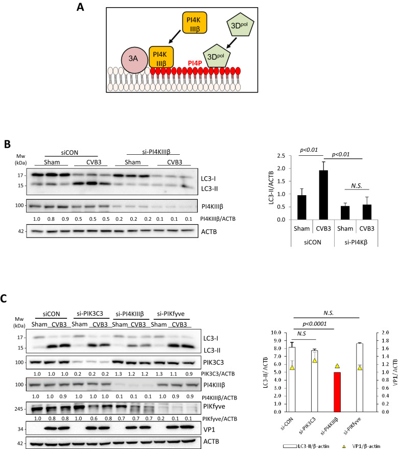
Figure 7.
PI4KIIIβ is involved in CVB3-induced LC3 lipidation. (A) Schematic diagram of the proposed role of PI4KIIIβ in autophagy and the known function in enterovirus replication. (B) PI4KIIIβ was transiently silenced in HEK293A cells using siRNA for 48 h. Cells were then subjected to sham or CVB3 infection for 16 h, followed by western blot analysis of LC3 and PI4KIIIβ. Densitometry was measured as above, and the results are presented underneath and in right panel (mean ± SD, n = 3. Analyzed by one way ANOVA with Tukey’s post-test). (C) HEK293A cells, transfected with control or PIK3C3, PI4KIIIβ, or PIKfyve siRNAs for 48 h, were sham- or CVB3-infected. Western blotting was performed for detection of LC3, VP1, PIK3C3, PI4KIIIβ, or PIKfyve, and quantified as above (right panel) (mean ± SD, n = 3. Analyzed by one way ANOVA with Tukey’s post-test). N.S. not significant.

