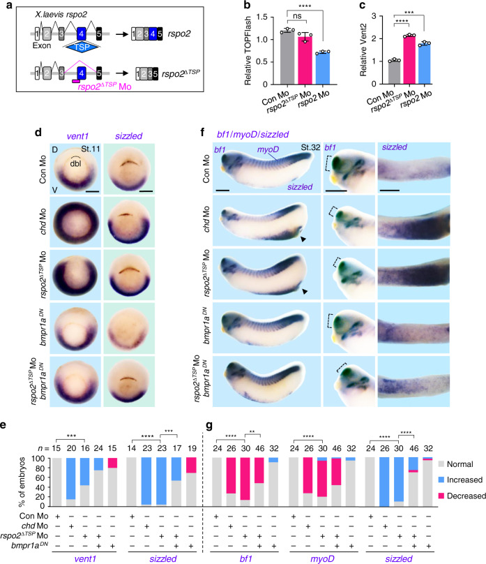
Fig. 5. Loss of Rspo2-TSP1 domain activates BMP signaling in Xenopus development.
a Scheme for rspo2∆TSP splicing Mo in Xenopus laevis. b TOPFlash assay in Xenopus laevis neurulae (St.15) injected radially at 4-cell stage with reporter plasmids and Mo as indicated. Data are displayed as mean ± SD; ns, not significant, ****P < 0.0001 from two-tailed unpaired t-test. n = 3 biologically independent samples. c BMP-reporter (vent2) assay in Xenopus laevis neurulae (St.15) injected radially at 4-cell stage with reporter plasmids and Mo as indicated. Data are displayed as mean ± SD; ***P < 0.001, ****P < 0.0001 from two-tailed unpaired t-test. n = 3 biologically independent samples. d–g In situ hybridization of BMP4 targets vent1 and sizzled in Xenopus laevis. Embryos were injected radially and equatorially at 4-cell stage as indicated. Gastrulae (St.11) (d) and quantification (e); Tadpoles (St. 32) (f) and quantification (g). Dashed lines, dorsal blastopore lip (dbl) (d) or bf1 expression (e); D, dorsal; V, ventral. For f, left, lateral view; middle, magnified view of head; right, magnified view of ventral side. ‘Increased/Decreased’ represents embryos with significant expansion/reduction of sizzled or vent1 signals toward the dorsal/ventral side of the embryo (e), or with significant increase/decrease of the signal strength (g). ns, not significant. n, number of embryos. Scale bar, 0.5 mm. Scoring of the embryos for quantification was executed with blinding from two individuals. For e, g, **P < 0.01, ***P < 0.001, ****P < 0.0001 from two-tailed χ2 test comparing normal versus increased. Data are pooled from at least two independent experiments.

