FIGURE 1.
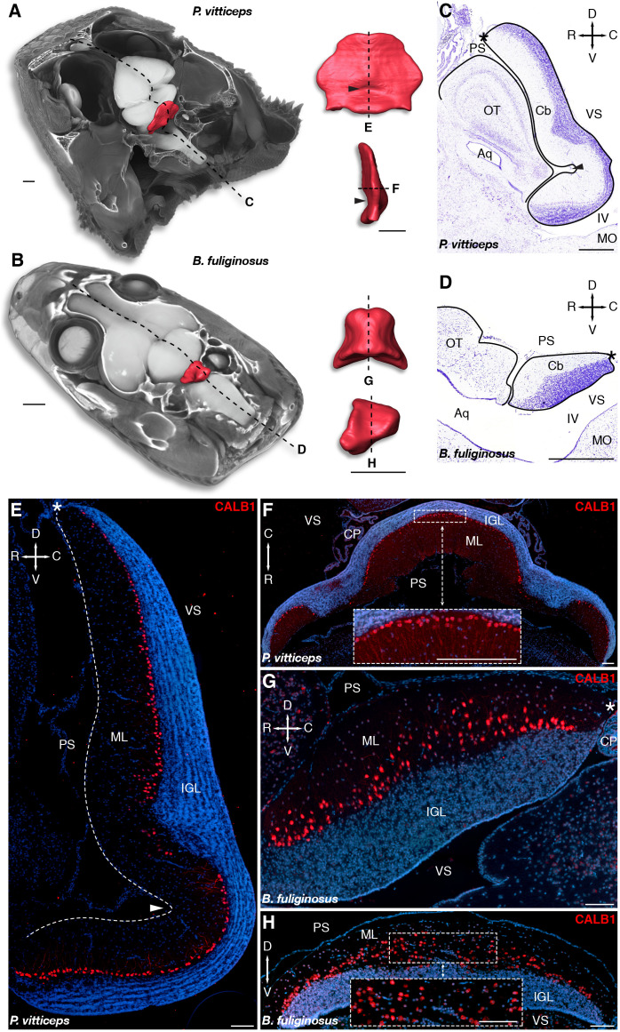
Morphology and cellular arrangement of bearded dragon lizard (P. vitticeps) and African house snake (B. fuliginosus) cerebellum. (A,B) 3D-volume rendering and high-resolution whole-brain segmentation of iodine-stained juvenile heads of P. vitticeps (A) and B. fuliginosus (B) highlighting the cerebellum structure (red color). High magnifications of 3D-rendered cerebella of P. vitticeps (A) and B. fuliginosus (B) are shown in pial (top) and lateral (bottom) views in the right column. Arrowheads indicate the position of the incomplete fissure in P. vitticeps cerebellum. Dashed lines and letters mark the sectioning planes relative to the histological preparations and immunostaining experiments in panels (C,D) and (E–H), respectively. (C,D) Nissl staining of the cerebellum and neighboring brain regions in P. vitticeps (C) and B. fuliginosus (D). Black lines demarcate the contour of the cerebellum and adjacent brain regions. The arrowhead in panel (C) indicates the position of the incomplete fissure in P. vitticeps cerebellum, and asterisks in panels (C,D) indicate the position of the embryonic upper rhombic lip. Crossed arrows point toward rostral (R), caudal (C), dorsal (D), and ventral (V) directions. (E–H) Immunodetection of Purkinje cells (PCs) with CALB1 marker, using sagittal (E) or axial (F) sections of P. vitticeps and sagittal (G) or coronal (H) sections of B. fuliginosus juvenile cerebellum (red staining). Cell nuclei are counterstained with DAPI (blue staining). The arrowhead in panel (E) indicates the position of the incomplete fissure, and the white dashed line delimitates the cerebellar pial surface. Asterisks in panels (E,G) indicate the position of the embryonic upper rhombic lip. Insets in panels (F,H) show high magnifications of PC spatial organization. Crossed white arrows point toward rostral (R), caudal (C), dorsal (D), and ventral (V) directions. OT, optic tectum; Cb, cerebellum; Aq, aqueduct; MO, medulla oblongata; IV, fourth ventricle; PS, pial surface; ML, molecular layer; IGL, internal granule layer; VS, ventricular surface; CP, choroid plexus. Scale bars: 1 mm (A,B), 500 μm (C,D), 100 μm (E–H).

