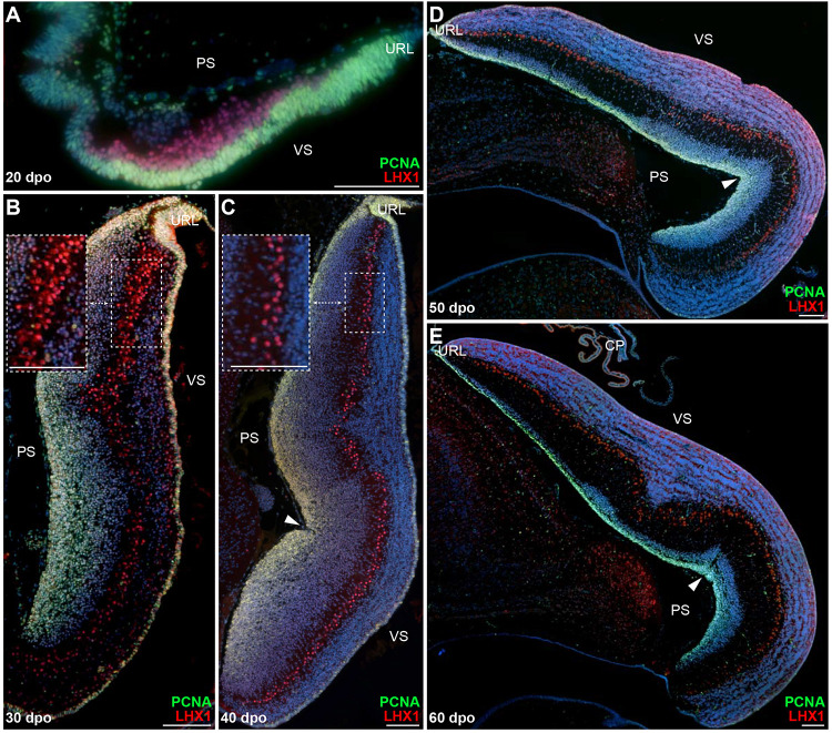
FIGURE 2.
Proliferation pattern and PC development in embryonic P. vitticeps cerebellum. (A–E) Double immunohistochemistry (IHC) staining for PCNA (green staining) and LHX1 (red) markers at various developmental stages, indicated as embryonic days post-oviposition (dpo), in the cerebellum of P. vitticeps. Arrowheads in panels (C–E) indicate the position of the incomplete fissure on the cerebellar pial surface. Insets in panels (B,C) show high magnifications of PC spatial organization. PS, pial surface; VS, ventricular surface; URL, upper rhombic lip. Scale bars: 100 μm.

