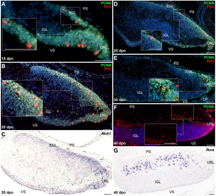
FIGURE 5.
Molecular characterization of GC patterning in B. fuliginosus. (A–E) Double IHC for PCNA (green staining) and PH3 (red) markers (A,B,D,E) or ISH for Atoh1 (C) at various indicated embryonic developmental stages between 15 and 30 dpo in the cerebellum of B. fuliginosus. Insets in panels (A,B,D,E) show high magnifications of mitotic progenitors on the pial surface. (F,G) Double IHC for PCNA (green) and SHH [(F); red] or ISH for Rora (G) at 40 dpo. The inset in panel (F) shows high magnification of SHH-positive PCs. PS, pial surface; EGL, external granule layer; IGL, internal granule layer; VS, ventricular surface; URL, upper rhombic lip; CP, choroid plexus. Scale bars: 50 μm.

