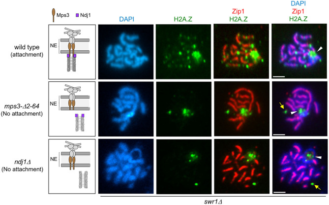
Figure 4.
H2A.Z localization at chromosome ends requires telomere attachment to the nuclear envelope (NE). Immunofluorescence of spread pachytene nuclei from the swr1Δ mutant stained with DAPI to visualize chromatin (blue), anti-GFP to detect H2A.Z (green), and anti-Zip1 to mark the synaptonemal complex (SC) central region (red). White arrowheads mark the ribosomal DNA (rDNA) region lacking Zip1. Yellow arrows point to H2A.Z foci likely corresponding to the spindle pole body (SPB; see text). The cartoons on the left schematize the linker of the nucleoskeleton and cytoskeleton (LINC) complex and the status of telomeric attachment in the different situations analyzed. Scale bar, 2 μm. The strains are DP1182 (wild type), DP1280 (mps3-Δ2-64), and DP1305 (ndj1Δ). Twenty-six, 28, and 24 nuclei were examined for wild type, mps3-Δ2-64, and ndj1Δ, respectively.

