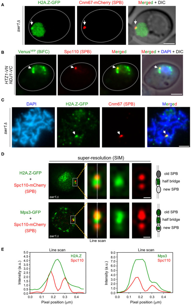
Figure 5.
H2A.Z localizes to the spindle pole body (SPB) half-bridge during meiotic prophase I. (A) Microscopy fluorescence image of a representative swr1Δ cell displaying a peripheral concentrated focus (arrow) of H2A.Z-GFP (green) colocalizing with the SPB marker Cnm67-mCherry (red). Images were taken from 16-h meiotic cultures. Scale bar, 2 μm. The strain is DP1172. (B) Bimolecular fluorescence complementation (BiFC) analysis of VenusYFP fluorescence (green) reconstituted from H2A.Z-VN/Ndj1-VC interaction in cells also expressing the SPB marker Spc110-RedStar2 (red). Nuclei were stained with DAPI (blue). A representative cell is shown. The arrow points to a single BiFC VenusYFP focus colocalizing with the SPB. Scale bar, 2 μm. The strain is DP1506. (C) Immunofluorescence of a spread pachytene representative nucleus stained with DAPI to visualize chromatin (blue), anti-GFP to detect H2A.Z (green), and anti-mCherry to mark the SPB (red). The arrow points to an H2A.Z focus colocalizing with Cnm67 (SPB). Scale bar, 2 μm. The strain is DP1172. Twenty-five nuclei were examined. (D) Structured illumination microscopy (SIM) fluorescence images of representative swr1Δ cells expressing Spc110-mCherry (red) and H2A.Z-GFP (top images) or Mps3-GFP (bottom images), in green. Scale bar, 0.1 μm. (E) Average intensity of the indicated proteins along the depicted line scan in all cells analyzed in (D). The strains are DP1578 (HTZ1-GFP SPC110-mCherry) and DP1576 (MPS3-GFP SPC110-mCherry); 33 and 26 cells were examined, respectively.

