
Figure 1.
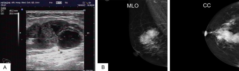
A. Ultrasonography demonstrated a hypoechoic mass in the right breast with multiple hypoechoic nodules around it, and the border was unclear. B. The mammography showed a high-density shadow in the upper quadrant of the right breast, and the edge was rough, measuring about 32 mm×23 mm in dimension (MLO position).