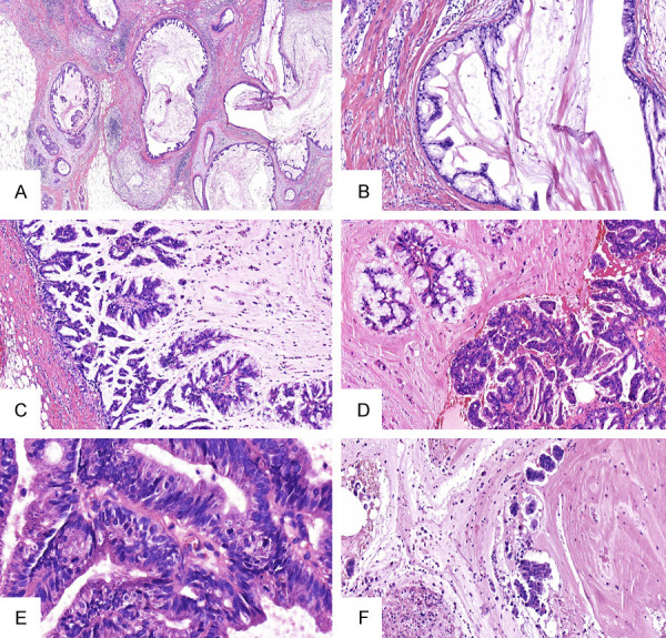
Figure 3.

A. Microscopically, the tumor showed variably sized cystic cavities lined by columnar cells and filled with hemorrhagic mucin (HE ×40). B. Some cavities are lined by monolayer high mild columnar cells with basally placed nuclei and abundant intracytoplasmic mucus (HE ×100). C. The cells in some regions were hyperplastic and protruded into the capsule cavity, forming a complex branching nipple (HE ×200). D. Some tumor cells have reduced mucus, increased atypia, and cytoplasmic eosinophilia (HE ×200). E. Pathological mitosis can be seen (HE ×400). F. A small amount of mucus overflows into the stroma, forming a mucus lake with the nested or papillary cell clusters floating there (HE ×100).
