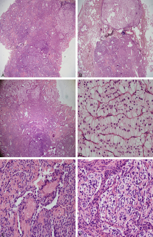
Figure 3.

A. The mass consisted of 3 parts (HE × 20). B. Another HE slice showed a lobulated mass (HE × 20). C. The mass consisted of 3 parts (HE × 40). D. The columnar clear cells area of the tumour (HE × 400). E. The classical sclerosing hemangioma-like area of the tumour (HE × 400). F. The short spindle solid cell nests area of the tumour (HE × 400).
