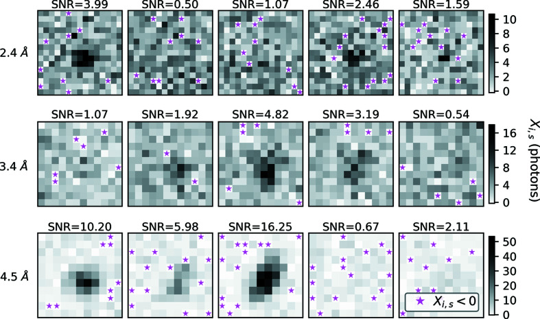
Figure 2.
Bragg spot profile variation observed in synthetic XFEL images. Each row contains five shoeboxes centered on the same Miller index, yet measured on separate shots (i.e. from different crystals). The resolution of each Miller index is shown in the far left of each row. In standard data analysis, integrated signals from shoeboxes with equivalent Miller indices are averaged together, leading to large uncertainties in the resulting structure factor estimates. Signal-to-noise ratio (SNR) estimates for the Bragg reflections inside each shoebox are provided for reference. We computed SNR following the work by Leslie (1999 ▸) (using a weighted least-squares treatment to propagate the tilt-plane error). Pixels with negative readings are marked with a star. The background scattering was very low in the synthetic data, as we attempted to accurately replicate in-vacuum diffraction from crystals in a 5 µm liquid jet produced by a GDVN (DePonte et al., 2008 ▸). Such low background in combination with per-shot readout errors gave rise to the negative pixels shown here. The gray scale represents X i,s (observed pixel intensity in units of photons) as defined in equation (8). All sub-images in a row are on the same scale (indicated by the scale bars on the right in units of photons). Note, the middle row appears to have fewer negative readings, which we expect because it is on the water scattering ring and, as a result, those pixels receive more background scattering. Fig. S1 shows the same set of images before random noise was applied.

