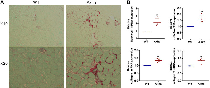
Fig. 2.
Renal interstitial fibrosis increases in 20-wk diabetic kidney disease mice. Renal interstitial fibrosis was estimated by sirius red staining. A: representative images of sirius red staining under microscopy with both ×10 and ×20 lens. Scale bars = 0.1 and 0.05 mm. B: quantitative analysis of fibronectin, α-smooth muscle actin (α-SMA), collagen type I, and collagen type IV by quantitative RT-PCR (n = 4). **P < 0.01 vs. the wild-type (WT) group.

