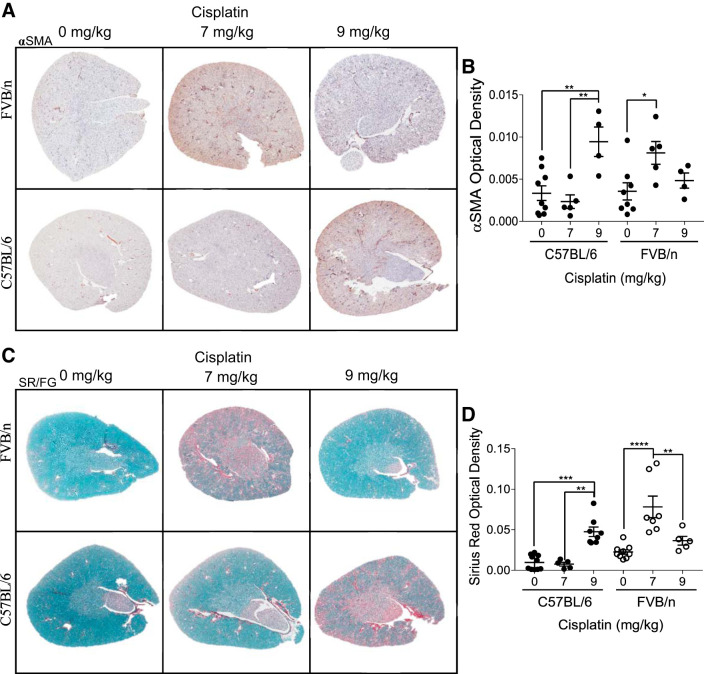
Fig. 2.
Development of renal fibrosis with repeated administration of cisplatin in C57BL/6 and FVB/n mice. A: α-smooth muscle actin (αSMA) immunohistochemistry for myofibroblasts. B: quantification of optical density of positive staining. C: Sirius Red/Fast Green (SR/FG) staining for total collagen deposition. D: quantification of optical density of positive staining. *P < 0.05, **P < 0.01, ***P < 0.001, and ****P < 0.0001.

