Fig. 3.
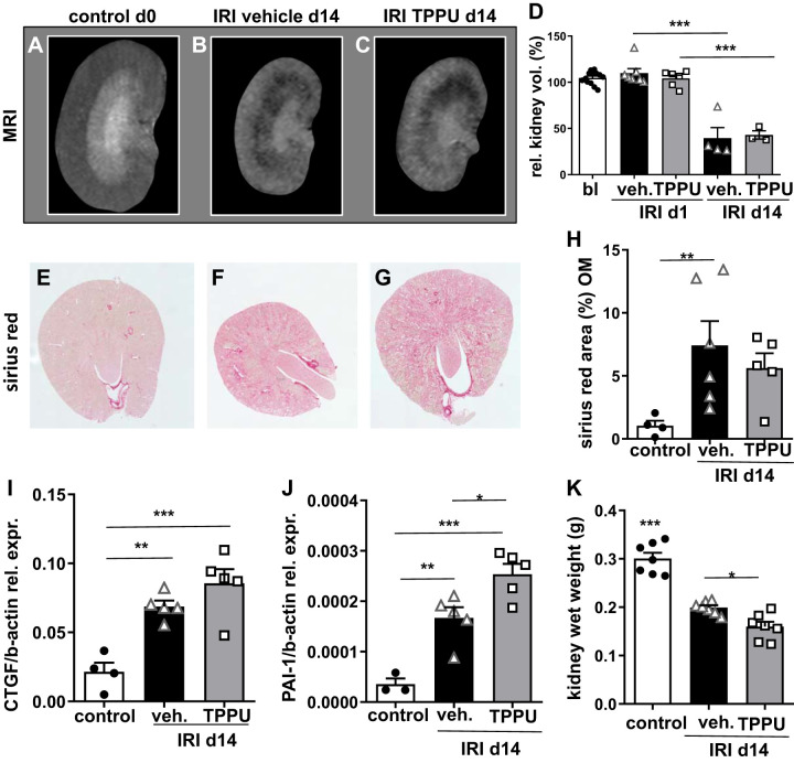
Kidney volume and chronic kidney disease at day 14 (d14) after ischemia-reperfusion injury (IRI). A−D: morphometric MRI imaging was done to assess kidney volume longitudinally and showed severe volume loss at day 14 after IRI in vehicle (veh)- and 1-trifluoromethoxyphenyl-3-(1-propionylpiperidin-4-yl) urea (TPPU)-treated mice. E−H: tubulointerstitial fibrosis showed enhanced collagen deposition after IRI in all groups (all images were kept in the same magnification and the stack function was used). Quantification of fibrosis in the outer medulla (OM) is shown. I: connective tissue growth factor (CTGF) mRNA was significantly increased in IRI kidneys compared with contralateral controls without differences between treatment strategies. J: plasminogen activator inhibitor-1 (PAI-1) mRNA expression was even higher in TPPU-treated compared with vehicle-treated IRI kidneys. K: the kidney wet weight decrease was more pronounced in TPPU-treated IRI kidneys compared with vehicle-treated kidneys. *P < 0.05; **P < 0.01; ***P < 0.001.

