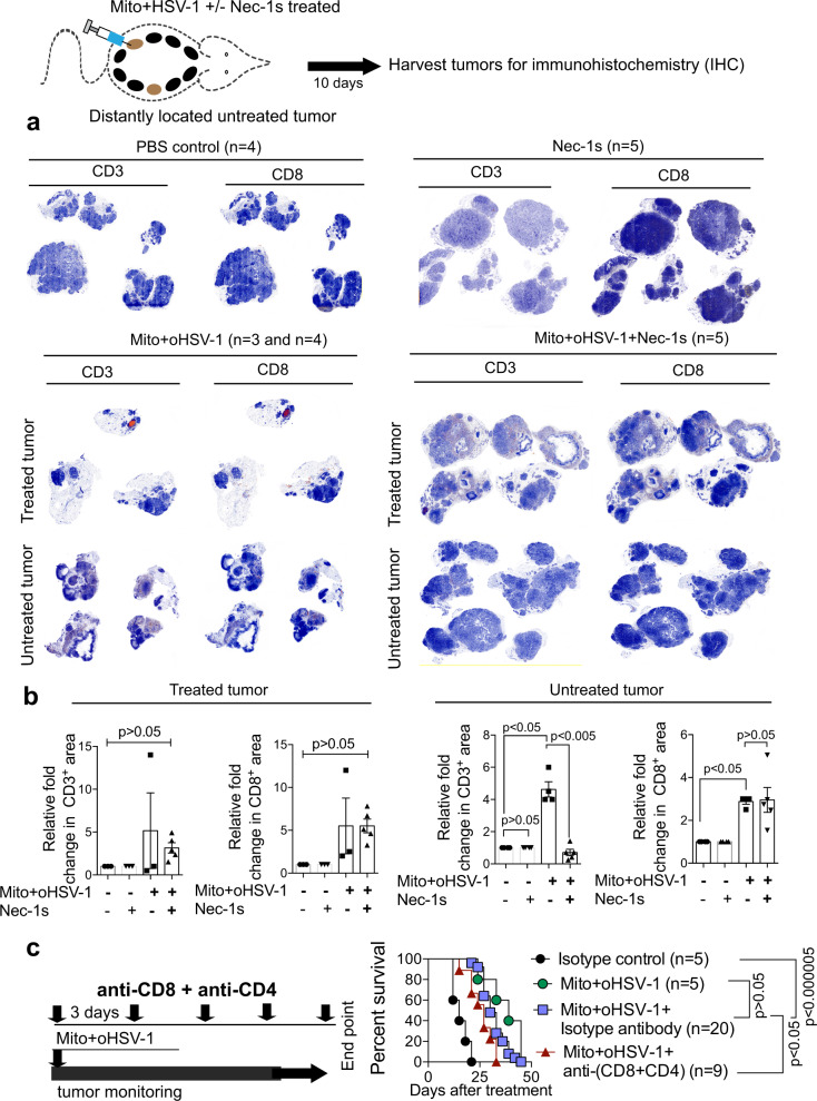
Fig. 5. Immunogenic Mito+oHSV-1 therapy CD8+ T-cell infiltration in distant tumor lesions.
a, b Ten days after Mito+oHSV-1 treatment treated and distant untreated tumor lesions were processed for IHC staining using CD3 and CD8 antibodies. Fold change in positive signal was calculated relative to the PBS controls. Quantitative data are mean±standard deviation from each treatment group (n = 5) (Statistical significance was tested with one-way ANOVA or non-parametric Kruskal–Wallis test with Dunn’s multiple comparison as a post hoc test). c BALB-neuT mice were treated with Mito+oHSV-1 along with monoclonal antibodies to deplete T cells (CD4 and CD8) or isotype control antibody. Quantitative data are Kaplan–Meier survival (Log-rank, Mantel–Cox test).

