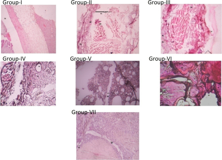
Figure 9.
Histological analysis of an ankle joint stained with H and E stain. A Gr. I-healthy control rat shows normal articular surfaces with a smooth layer of cartilage (hyaline). A Gr. II-FCA-induced adjuvant arthritic rat showed (a) massive cell infiltration, (b) synovium, (c) joint cavity with a large joint space, (d) pannus formation, and (e) hyperanemia with dilated blood vessels. A Gr. III-FCA rat treated with the blank liposome showed (a) degradation of the articular surface and (b) infiltration of inflammatory cells. Gr. IV-in FCA rat treated with a combination of free drug NAR + SFN, (a) cellular infiltration is still present, which is composed of neutrophils representing acute inflammation and (b) vacuolar degeneration is also seen. Gr. V-in the synovial tissues treated with NAR + SFN CLF, fewer leukocytes were present and showed normal articular cartilage with (a) lesser joint space. Gr. VI-FCA rat treated with free drug combination of NAR + PEITC showed the presence of (a) cellular infiltration and (b) vacuolar degeneration. Gr. VII-adjuvant rat treated with NAR + PEITCCLF showing reduction of cell infiltration with (a) decrease in joint.

