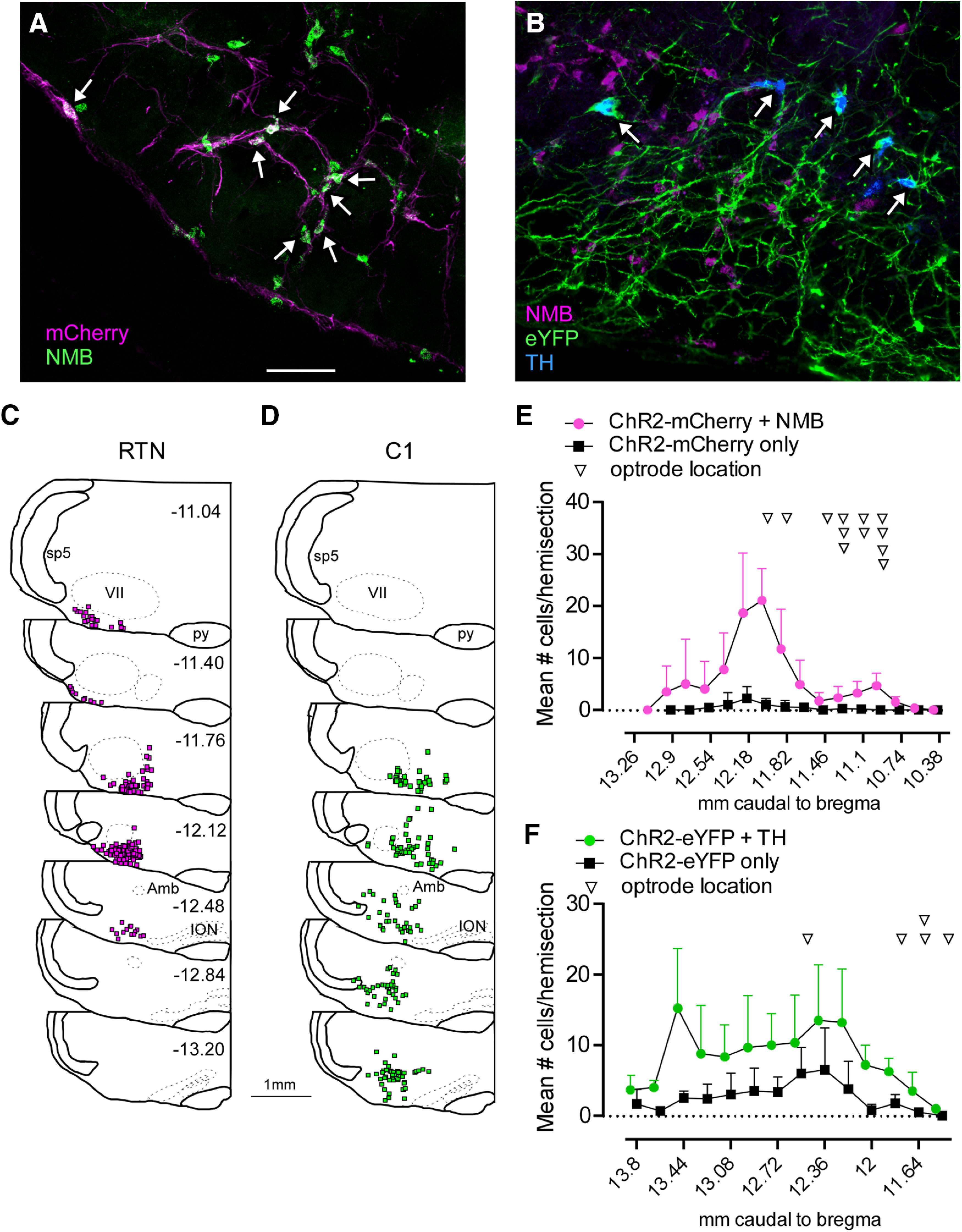
Figure 1.

Selective expression of ChR2 in RTN and C1 neurons. A, Combined ISH and immunohistochemistry showing selective transduction of RTN neurons (Nmb+mCherry, white arrows) in a Th-Cre rat in which Th neurons were ablated by injecting rAVV5-Flex-taCaspase3-Tevp (transverse section; left). B, ISH showing the selective transduction of C1 neurons (Th+eYFP, white arrows). C, Rostral-to-caudal series of transverse sections (bregma levels in mm as indicated) depicting the location of Nmb+ChR2-mCherry-expressing neurons overlaid from 3 RTN-targeted cases. D, Rostral-to-caudal series of transverse sections depicting the location of Th+ChR2-eYFP-expressing neurons overlaid from 3 C1-targeted cases. E, Neuronal distribution and optical fiber location across bregma levels in RTN-targeted cases. F, Neuronal distribution and optical fiber location across bregma levels in C1-targeted cases. References to mm caudal to bregma after Paxinos and Watson (2014). Scale bar, 100 µm.
