Figure 1.
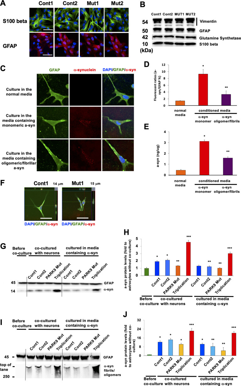
Astrocytes absorb different α-syn species from culture media. A, Astrocytes differentiated from iPSCs that were taken from two normal individuals (Cont 1 and Cont 2) and two patients with ATP13A2 mutations (Mut 1 and Mut 2). The cells were stained with astrocyte markers GFAP (top) or β-S100 (bottom). B, Immunoblot characterization of astrocytes with astrocyte markers including vimentin (top), GFAP (second from top), glutamine synthetase (second from bottom), and β-S100 (bottom). C–E, iPSC-derived astrocytes can absorb α-syn from the media. C, Representative images of α-syn immunofluorescence in Cont 1 astrocytes before (top) and after culturing in the media containing monomeric α-syn (middle) and oligomeric/fibrillar α-syn (bottom) for 24 h. GFAP is used for astrocytic marker. D, Quantification of α-syn fluorescence intensities normalized by a nuclear marker, 4′,6′-diamidino-2-phenylindole dihydrochloride (DAPI) intensities in astrocytes (n = 3, *p = 0.001, **p = 0.015, one-way ANOVA with Tukey's post hoc test). E, The quantification of α-syn levels in astrocytes that were cultured in the normal media or in the media containing α-syn (conditioned media; n = 3, *p = 0.001, **p = 0.0011, one-way ANOVA with Tukey's post hoc test). F, Trypan blue assay with Cont 1 and Mut 1 astrocytes that were cultured in the media containing oligomeric/fibrillar α-syn for 24 h. G, Representative immunoblot for α-syn of lysates from astrocytes before (leftmost lane), after being cocultured with Cont 1 and 2 and Mut 1 DA neurons and DA neurons harboring α-syn triplication (lanes 2–5), and after being cultured in the leftover media of Cont 1 and 2 and Mut 1 DA neurons and DA neurons harboring α-syn triplication (lanes 6–9). H, Densitometric quantification is shown as the relative α-syn levels against GFAP in astrocytes (n = 3, *p = 0.003, **p = 0.042, ***p = 0.001, one-way ANOVA with Tukey's post hoc test). I, Representative immunoblot for α-syn of lysates from astrocytes before (leftmost lane), after being cocultured with Cont 1 and 2 and Mut 1 DA neurons and DA neurons harboring α-syn triplication that were cultured in the media containing oligomeric/fibrillar α-syn for 24 h (lanes 2–5) or cultured in the leftover media of Cont 1 and 2, and Mut 1 DA neurons and DA neurons harboring α-syn triplication after they were cultured in the media containing oligomeric/fibrillar α-syn for 24 h (lanes 6–9). J, Densitometric quantification is shown as the relative α-syn levels against GFAP in astrocytes (n = 3, *p = 0.001, **p = 0.0013, ***p = 0.003, one-way ANOVA with Tukey's post hoc test). Scale bars: A, C, and F, 50 µm. In all graphs, error bars indicate the SEM.

