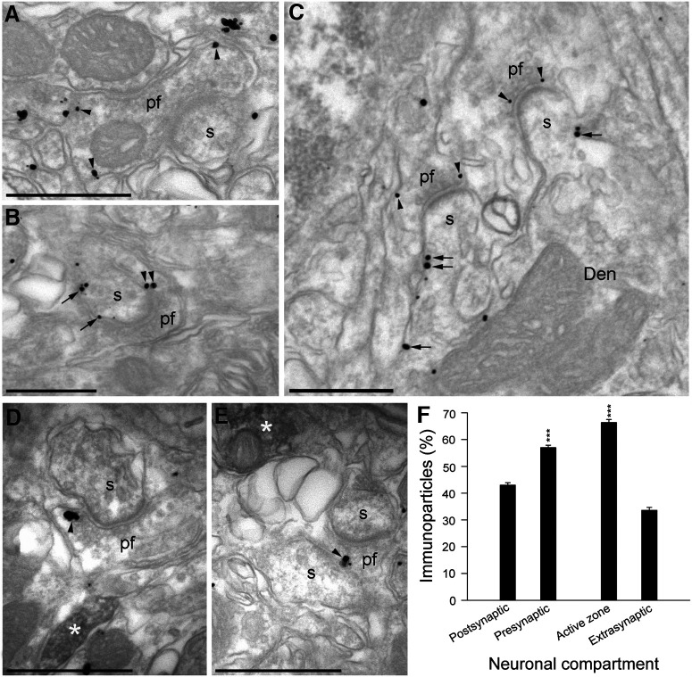
Figure 3.
Subcellular localization of the β1-adrenergic receptors in the presynaptic compartments of the cerebellum. A–C, Electron micrographs of the molecular layer of the cerebellum showing pre-embedding Immunogold staining for β1AR. Immunoparticles for β1AR were observed along the plasma membrane (arrowheads) of parallel fiber terminals (pf) establishing excitatory synapses with dendritic spines (s) of PCs. Less frequently, immunoparticles for β1AR were also observed at postsynaptic sites along the plasma membrane (arrows) of dendritic spines (s) and the shafts (Den) of PCs. D, E, Electron micrographs of the molecular layer of the cerebellum showing β1AR immunoparticles and the immunoperoxidase reaction for TH detected using a dual-labeling pre-embedding method. β1AR immunoparticles were observed along the plasma membrane (arrowheads) of pf terminals, always close to fibers immunopositive for TH (filled with the peroxidase reaction end product, white asterisks). F, Quantitative analysis showing the percentage of β1AR immunoparticles in the molecular layer of the cerebellum. Immunoparticles (502) for β1ARs were more frequently observed in presynaptic compartments (57.0 ± 0.9%) and within axon terminals than in the postsynaptic compartments (43.0 ± 0.9%, t = 10.999, df = 4). Moreover, they were more frequently found in the active zone (66.4 ± 1.1%, t = 21.085, df = 4) than at extrasynaptic sites (33.6 ± 1.1%). Scale bars: A, D, E, 500 nm; B, C, 200 nm. The data represent the mean ± SEM.

