
FIGURE 4.
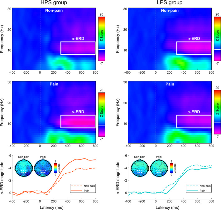
Time‐frequency responses during the Pain Judgment Task. Time‐frequency distributions of neural responses for HPS and LPS groups were elicited by nonpainful and painful stimulations during the Pain Judgment Task. The color scale represents the increase or decrease of the oscillatory magnitude relative to a prestimulus interval (−400 to −100 ms). Displayed signals were measured at centro‐parietal electrodes (CP1, CPz, CP2, P1, Pz, and P2). Both nonpainful and painful stimulations elicited a long‐lasting α‐ERD response (8–14 Hz in frequency and 300–800 ms in latency, marked using white rectangles). The time‐course of α‐ERD magnitudes in response to painful (solid line) and nonpainful (dashed line) stimulations was obtained by averaging across 8–14 Hz. Electrodes used to evaluate the α‐ERD magnitude are marked using enlarged white dots on the scalp topographies