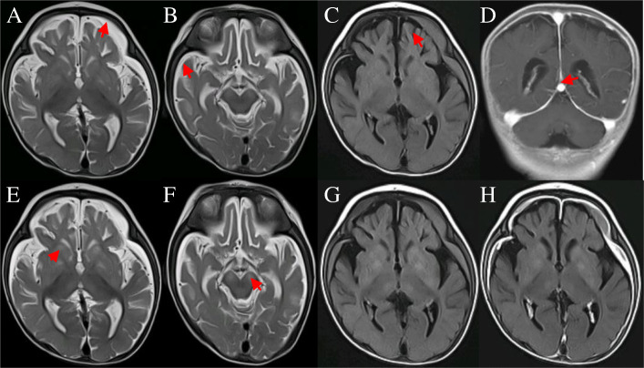
Fig. 1.
3T Brain MRI of the patient: a to d were performed when he was first transfer to our hospital at 4-month-old. a and b: axial T2WI showed arc-shaped subdural empyema was seen in bilateral frontotemporal parietal lobes. c: axial T2 Flair showed arc-shaped subdural empyema and bilateral cerebral hemisphere atrophy. d: Coronal T1WI enhanced scanning showed bilateral frontotemporal parietal dura enhanced which was called Mercedes-Benz sign. e to h was reexamined 7 days after hospitalization. e and f: axial T2WI showed new symmetrical hypersignal lesions in bilateral basal ganglia, thalamus and cerebral peduncle. g: the lesions had a slightly higher signal on T2Flair. h: the lesions had no obvious enhancement on T2Flair

