Fig. 6.
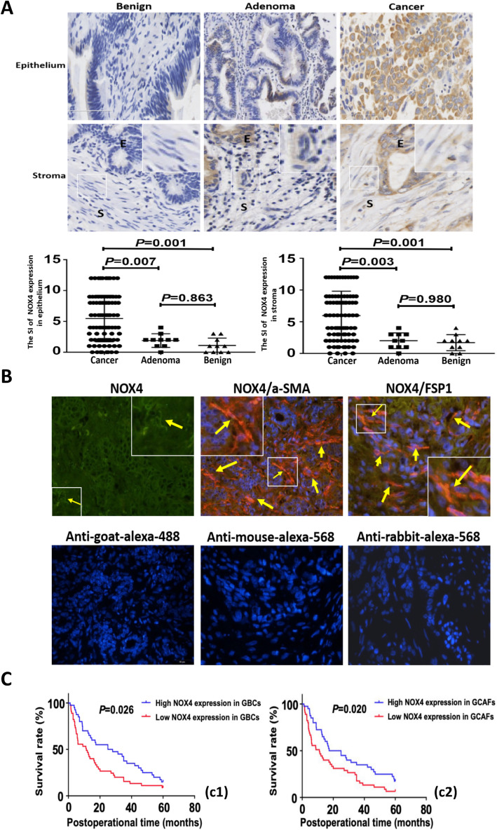
NOX4 is highly expressed in human GBC tissue/stroma, and is associated with poor prognosis. A (IHC, × 200; E = epithelium, S=Stroma). The expression (cytoplasmic and/or nuclear brown staining) of NOX4 in epithelium or stroma of GBC (n = 85) was significantly upregulated compared with gallbladder precancerous (adenomas, n = 10; epithelium: 5.471 ± 0.410 vs. 1.900 ± 0.348, P = 0.007, stroma: 5.965 ± 0.419 vs. 2.000 ± 0.394, P = 0.003) and benign lesions (cholecystitis, n = 10; epithelium: 5.471 ± 0.410 vs. 1.100 ± 0.379; stroma: 5.965 ± 0.419 vs. 1.700 ± 0.396; both P = 0.001), but no statistical difference between precancerous lesions and benign lesions (both P > 0.05). Magnified insets show representative NOX4 staining in stroma. B (CIF staining, × 200). The expression of NOX4 (green) co-localizes with α-SMA and FSP1 (red) positive stroma. Representative samples of NOX4, α-SMA and FSP1 are shown. Sections were counterstained with DAPI. Secondary antibody only controls are shown: anti-goat-alexa-488 for NOX4, anti-mouse-alexa-568 for α-SMA and anti-rabbit-alexa-568 for FSP1. Arrows and inset point to positive staining in fibroblastic cells. C. Kaplan-Meier analysis of GBC patients with high and low NOX4 expression in GBC cells/stroma. The survival time of GBC patients with high NOX4 expression in tumor cells or stroma was significantly shorter than that of GBC patients with low NOX4 expression (log-rank test, c1, GBCs; P = 0.026; c2, GCAFs, P = 0.020)

