Figure 1.
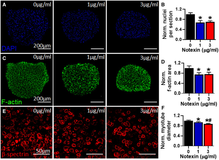
Effects of notexin injury on myobundle structure. A, Representative myobundle cross-sectional images stained for nuclei (DAPI). B, Quantification of nuclei number per myobundle cross-section normalized to drug-free control (n = 4 myobundles per group, n = 2 technical replicates from N = 1 donor). C, Representative myobundle cross-sectional images stained for filamentous actin (f-actin). D, Quantification of F-actin positive area per myobundle cross-section normalized to drug-free control (n = 4 myobundles per group from N = 1 donor). E, Representative myobundle cross-sectional images stained for β-spectrin. F, Quantification of myotube diameter relative to drug-free control (n = 100 myotubes from 4 myobundles per group from N = 1 donor). *p < .01 from 0 μg/ml notexin and #p < .01 from 1 μg/ml notexin.

