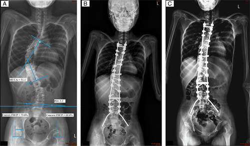
FIGURE 1.

All the posterior-anterior radiography was made with the patient in supine position with the head, trunk, and lower extremities as anatomically straight as possible. A, Preoperative radiography of a 12-year-old girl with closed triradiate cartilage and a 51.1 scoliosis. The major curve of Cobb angle (MCCA), pelvic obliquity (PO), and femoral head coverage percentage (FHCP) were measured as previously described.4,15,16 B, The patient had received segmental spinal instrumentation with pedicle screws and the Galveston pelvic fixation technique. The fusion level was from T2 to the pelvis. Postoperative radiography showed that the residual MCCA was 21.1 degrees and the residual PO was 2 degrees. C, Postoperative radiography at 24 months, with no obvious change in MCCA or PO. “Windshield wiper” sign (iliac bone osteolysis) was observed but this did not cause fusion defects or decrease the PO correction.
