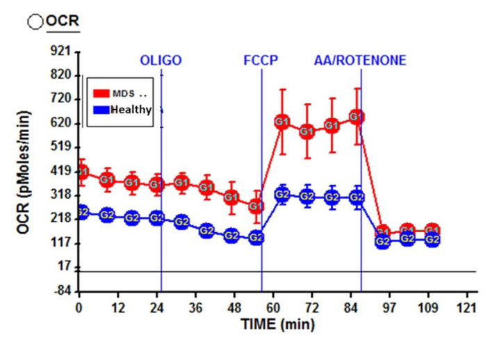
Figure 3. Increased basal and maximal respiratory capacity in an MDS patient with excess of blasts.
OCR measurements of peripheral blood cells obtained from an MDS patient (Red) in comparison to his matched healthy control (blue). Each dot represents the average of 3 repeats. Cells were plated on a Cell-Tak™ coated 24 well XF V7 cell culture microplate at 0.5×10⁶ cells per well in 50 μL of XF assay medium.

