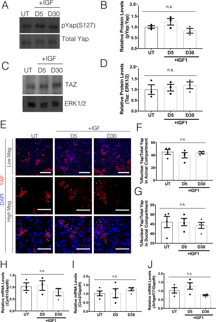
Fig 2. Post-therapeutic IGF1 treatment reduces Yap activation.
FVB mice (4–6 weeks old) were either untreated (UT) or irradiated (IR) with 5Gy and given post-therapeutic IGF1 (IR+IGF) on days 4–7. Parotid salivary glands were dissected on days 5 and 30 following radiation treatment. Levels of (A-B) phosphorylated Yap on serine 127 (pYap S127) and (C-D) Taz were evaluated following IR+IGF treatment. Immunoblots were re-probed with total Yap or ERK as a loading control. (E) Immunofluorescent staining was used to determine the percentage of cells that displayed nuclear Yap (red) in the acinar compartment. Composite images with DAPI (blue) are presented in both high and low magnification (scale bar for high magnification = 30 μm, low magnification = 100 μm). Quantification of the percentage of Yap positive cells with nuclear Yap staining from E was determined in the acinar enriched (F) and ductal (G) compartments separately. Relative mRNA levels of Cyr61 (H), Ccn2 (I) and Arhgef17 (J) were determined by qRT-PCR and normalized to Gapdh. Results are presented from at least four mice per condition (each data point graphed in B, D, F-J represents individual mice); E-F used 10–15 images per mouse; error bars denote mean ± SEM. Multiple comparisons were conducted using a Tukey-Kramer test and significant differences (p<0.05) between treatment groups are denoted with different letters above the bar graphs. Treatment groups with different letters are significantly different from each other.

