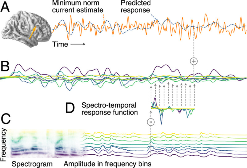
Fig 1. Additive linear response model based on STRFs.

(A) MEG responses recorded during stimulus presentation were source localized with distributed minimum norm current estimates. A single virtual source dipole is shown for illustration, with its physiologically measured response and the response prediction of a model. Model quality was assessed by the correlation between the measured and the predicted response. (B) The model’s predicted response is the sum of tonotopically separate response contributions generated by convolving the stimulus envelope at each frequency (C) with the estimated TRF of the corresponding frequency (D). TRFs quantify the influence of a predictor variable on the response at different time lags. The stimulus envelopes at different frequencies can be considered a collection of parallel predictor variables, as shown here by the gammatone spectrogram (8 spectral bins); the corresponding TRFs as a group constitute the STRF. Physiologically, the component responses (B) can be thought of as corresponding to responses in neural subpopulations with different frequency tuning, with MEG recording the sum of those currents. MEG, magnetoencephalographic; STRF, spectrotemporal response function; TRF, temporal response function.
