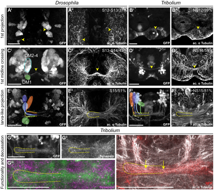
Fig 5. Key events of central complex development occur during late embryogenesis in Tribolium but not in Drosophila.
Comparable steps of central complex development are shown in one row for both species, the respective stage/relative time of development are shown in panels ii. The analysis was based on EGFP-labeled neurons (panels i), whereas acetylated α-tubulin staining is shown for reference (panels ii). (A-B) The development of the first axons happened at a similar time in Drosophila and Tribolium. (C-D) First midline-crossing fibers appeared earlier in Drosophila. (E-F) Likewise, the larva-like projection pattern was reached earlier in Drosophila. (G-H) The late-stage embryonic central complex of Tribolium is already faintly synapsin-positive (Gii, magenta in Giii), whereas the Drosophila lvCB remains synapsin-negative. In Tribolium, first decussations were visible (H, yellow arrows). Note that the assignment of rx-positive cell clusters to the DM1-4 lineage groups was not unambiguous before midembryogenesis. Tentatively, we indicated the location of DM1 (green) and DM2-4 cells (blue oval form) in Ci. Later, the groups could be assigned to DM1-4 lineages (E-F). Stages in Drosophila correspond to [68] and in Tribolium to [37]. Posterior is up, except in panels F, G, and H where dorsal is up. Scale bars represent 25 μm. ac, anterior commissure; GFP, green fluorescent protein; lvCB, larval central body; NS, neural stage; Rx, retinal homeobox. Original data: https://figshare.com/articles/Fig5/11339810.

